Νέα 2026
New treatments for severe autoimmune diseases
- Written by: Dr. Kouzi
Immune therapies in the treatment of autoimmune diseases
Results announced in EULAR 2026 Congress
A. Dual-target CAR T-cell therapy exhibits ‘profound’ remission in systemic sclerosis (SSc)
- All 11 patients achieved rapid B-cell aplasia, with significantly improved skin thickness scores, while 73% achieved low disease activity.
- The immune “reset” strategy has demonstrated “curative potential,” the researchers wrote.
A dual-target CD19/BCMA CAR T-cell therapy yielded “profound, sustained” remission in 11 patients with refractory systemic sclerosis, according to data presented at the EULAR 2026 Congress. “As we all know, despite advances in immunosuppressive, biologic and antifibrotic therapies, a subset of patients with refractory, progressive systemic sclerosis continues to experience uncontrolled skin fibrosis, lung involvement and functional decline,” Yajing Zhang, MD, PhD, of Beijing GoBroad Boren Hospital, in China, told attendees at the meeting. “CD19 targeting depletes pathogenic B-lineage cells, whereas BCMA targeting may extend immune reset toward plasma-cell-associated autoantibody production, a compartment potentially relevant to refractory SSc.” To examine the efficacy and safety of dual-target CD19/BCMA CAR T-cell therapy in refractory SSc, including the potential for remission induction, Zhang and colleagues recruited a total of 11 patients. Participants had a median disease duration of 4 years and had previously received immunosuppression with cyclophosphamide and rituximab (Rituxan, Genentech). Among the participants, nine demonstrated diffuse cutaneous SSc and five had interstitial lung disease at baseline. Following lymphodepletion, all 11 participants received autologous CD19/BCMA CAR-T cells, at a dose of 1 to 2.2×10⁶/kg and a high-resolution CT at baseline and monthly up to month 6 of follow-up. According to the researchers, all 11 patients demonstrated rapid B-cell aplasia, defined as a CD19+ rate of less than 0.1%, via flow cytometry, at day 15 to month 1. In all, 73% of participants achieved an mRSS of less than 10, denoting low disease activity. Additionally, mRSS “improved significantly” from a baseline median of 19 to a median of 4 at month 3 (P = .002), and a median of 2 at month 6 (P < .001), the researchers wrote.
Regarding lung involvement, patients demonstrated forced vital capacity/diffusing capacity of the lungs for carbon monoxide stabilization or improvement. Lastly, 80% of patients demonstrated regression of interstitial changes on high-resolution CT. No disease flares occurred during a median 4 months of follow-up, according to the researchers. One patient with overlap syndrome “required retreatment but achieved remission post-second infusion,” they wrote. “Dual-target CD19/BCMA CAR-T cell therapy induces profound and sustained clinical remission in refractory systemic sclerosis,” Zhang said in a EULAR press release announcing the data. “By effectively targeting both skin fibrosis and lung progression, this immunological ‘reset’ strategy offers true curative potential, paving the way for phase 2 trials to redefine the future management of this severe disease.” The study from Zhang’s group was one of several presented at the EULAR 2026 Congress highlighting CAR T-cell therapies across a host of autoimmune rheumatic diseases.
Β. Early remission in rheumatoid arthritis with CAR T
Fredrik N. Albach, MD, of Charité-University Medicine Berlin, presented data from the phase 1, prospective, open-label COMPARE trial, which assessed CAR T in six patients with refractory active rheumatoid arthritis. According to the researchers, the treatment, a fully human CD19-directed CAR-T cell therapy, was well tolerated, with mild-to-moderate cytokine release syndrome reported in three patients. After treatment, CAR T cells expanded rapidly and peaked at a median of 227.7 cells/µL within 3 weeks, followed by a gradual decline. B cells were “profoundly” depleted in blood and tissues among all patients, the researchers wrote. Albach and colleagues noted a “marked decrease” in autoantibodies — a greater than 90% median reduction — as well as sustained seroconversion to normal ACPA and RF-IgM achieved in four and five patients, respectively, according to a EULAR press release. All participants demonstrated decreased disease activity, with median 49% DAS28-CRP reduction and ACR20/50/70 responses in five, four and two patients, respectively. Additionally, three patients achieved sustained, immunosuppressive-free remission.
C.CAR T’s impact on the gut microbiome
According to Yuichi Maeda, MD, of Friedrich-Alexander University of Erlangen-Nuremberg, in Germany, and colleagues, CD19 CAR T-cell therapy “reshapes” gut microbiota in patients with autoimmune inflammatory disease by reducing Bifidobacterium adolescentis. Fecal IgA and serum anticommensal IgA and IgG responses also decreased.
The researchers noted that B. adolescentis “has been shown to promote intestinal Th17 responses, exacerbate experimental arthritis and impair epithelial barrier function.” “These immunomicrobial shifts may support long-term disease remission,” Maeda and colleagues wrote. “Notably, lower baseline fecal IgA predicted delayed B-cell reconstitution at 6 months, highlighting mucosal immune status as a potential regulator of post CAR T-cell immune recovery.” The researchers assessed participants’ gut microbiota and fecal IgA levels prior to and 6 months after treatment.
D.Novel E-CAR induces remission in SLE, SSc
To overcome the toxicities of the using of conventional CAR T, as an alternative scientists in China use a novel construct called E-CAR. A poster presented by Xiaobing Wang, MD, of the National Key Laboratory for Immunity and Inflammation, at Shanghai Changzheng Hospital, revealed data from a first-in-human pilot study of E-CAR T cells in three patients with SLE and one with SSc. According to the researchers, the patient with SSc demonstrated “dramatic fibrosis reversal and resolution of interstitial lung disease.” Meanwhile, the patients with SLE demonstrated complete clearance of immune complex deposition vs. baseline, confirmed by repeated renal biopsies at month 10. “This pilot study demonstrates that physiological signaling via the CD3-based E-CAR drives deep, tissue-level remission with a superior safety profile compared to conventional designs,” Wang and colleagues wrote. “The differentiation of E-CAR T cells into a TRM phenotype provides a novel mechanism for sustained disease control despite peripheral clearance.”
The CEO of Biohellenika is in the top 2% of the world scientists for the year 2023
- Written by: Dr. Kouzi
Biohellenika staff congratulates the CEO, founder and visionary of the company, Professor Emeritus of Biochemistry at the Medical School of the Aristotle University of Thessaloniki, Mr. Georgios Koliakos, for the global recognition of his scientific work in the field of Medicine and Biology and his ranking in the top 2% of scientists in the world, according to the published list of the University of Stanford.
The ranking is conducted based on the size and significance of his published work during the year 2023, and concerns the evaluation of more than 6,000,000 scientists worldwide. The work of these scientists has accelerated progress in their respective fields of research and has influenced the research and career of other scientists.
Even 10 years after allogeneic hematopoietic transplantation patients suffer from serious side effects
- Written by: Dr. Kouzi
Late GVHD remains common and lethal up to 10 years after allogeneic hematopoietic transplantation
Late acute and chronic graft vs host disease (GVHD) remain frequent complications and continue to drive long-term mortality following allogeneic hematopoietic cell transplantation (HCT), according to a 10-year follow-up study from the Chronic GVHD Consortium published in Transplantation and Cellular Therapy.
The investigators conducted a long-term follow-up analysis of a prospective, multicenter cohort study to characterize the incidence, clinical course, and outcomes of late acute GVHD, chronic GVHD, and key chronic GVHD subtypes over an extended follow-up period.
The study included 911 patients undergoing first allogeneic HCT who were enrolled pretransplant or within 4 months post-transplant between 2011 and 2014 and followed for up to 10 years. Patients were assessed longitudinally for development of GVHD syndromes, organ involvement, treatment patterns, and survival outcomes.
The cohort had a median age of 53.7 years (range, 19-78 years), 61% were male, and most had acute leukemia or myelodysplastic syndromes (63%). Peripheral blood grafts were used in 80% of patients, and most (76%) received matched related or unrelated donor transplants.
Over long-term follow-up, late acute GVHD developed in 11% of patients, with a median of 5.5 months after HCT. Chronic GVHD developed in 54% of patients, with a median onset of 7.4 months. Less common but more severe manifestations included BOS (4%) and cutaneous sclerosis (10%), both of which presented later, at medians of 12.6 and 17.2 months, respectively.
The analysis revealed that new GVHD events continued to occur beyond the first 1 to 2 years post-transplant, underscoring the prolonged risk period. Interconversion between GVHD subtypes was common; for example, 46% of patients with late acute GVHD subsequently developed chronic GVHD, and a subset of patients with chronic GVHD later developed bronchiolitis obliterans syndrome (5%) or cutaneous sclerosis (13%).
The investigators reported that clinical outcomes remained poor across GVHD subtypes. Nonrelapse mortality increased over time, reaching 35% at 10 years among patients with late acute GVHD and 31% among those with chronic GVHD, with particularly high mortality observed among patients with bronchiolitis obliterans syndrome (62%). Overall survival at 10 years after GVHD onset was 53% among patients with late acute GVHD, 54% among those with chronic GVHD, and only 28% among those with BOS.
This long-term analysis provides extensive coverage on incidence, presenting features, and outcomes following late acute GVHD, chronic GVHD, cutaneous sclerosis, and bronchiolitis obliterans syndrome after HCT, and supports the need for late post-HCT monitoring for these syndromes.
Durable discontinuation of systemic immunosuppression was uncommon. Approximately 40% of patients achieved complete discontinuation by 5 years after diagnosis for most GVHD subtypes, with lower rates observed in BOS (20%).
At a population level, GVHD-free, relapse-free survival remained low, declining from 22% at 2 years to 15% at 10 years post-transplant.
“In summary, this long-term analysis provides extensive coverage on incidence, presenting features, and outcomes following late acute GVHD, chronic GVHD, cutaneous sclerosis, and BOS after HCT, and supports the need for late post-hematopoietic stem cell transplantation monitoring for these syndromes,” the study authors concluded in their report. “The data support the importance and impact of established post hematopoietic transplantation late acute and chronic GVHD in several ways, notably including the adverse impact of these syndromes on mortality and prolonged requirements for immune suppressive therapy.”
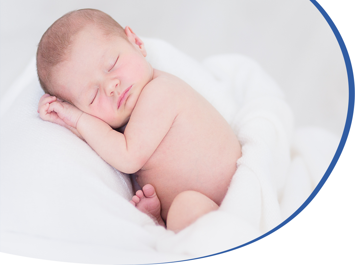
The above very important findings are complications of allogeneic transplantations, in which hematopoietic stem cells from compatible donors are used and justify the need for new safer and less traumatic treatments for malignant diseases. The hematopoietic grafts used are obtained from umbilical cord blood, bone marrow and peripheral blood. Cord blood stem cells, due to their very young age, have not yet expressed the markers of histocompatibility, they are better compatible and are characterized by lower rejection rates. Due to their limited number, hematopoiesis is delayed, but after this stage the patient's course is safer.
Transplantations are characterized by serious complications either due to high doses of chemotherapy or due to long-term use of immunosuppressants.
For these reasons, in recent years, cell therapies and immunotherapies have been developed, which are characterized by better outcomes and much fewer side effects. These therapies are autologous, created by the patient's own T lymphocytes, the chemotherapy is only introductory, immunosuppressants are not used and are applied even at very old ages. For these reasons, they have now replaced 50% of classic transplantations. T lymphocytes are present in umbilical cord blood, they are healthy, they are in a healthy environment and are collected together with hematopoietic stem cells. The T lymphocytes from a patient suffering from a malignant disease are mixed with malignant cells. Therefore, umbilical cord blood is in many ways a safe transplant for any type of treatment, autologous or allogeneic, classic or innovative.
Pidala J, Onstad L, Carpenter P, et al. Longitudinal study of late acute and chronic graft-versus-host disease after allogeneic hematopoietic cell transplantation: a long-term follow-up study from the chronic graft-versus-host disease consortium. Transplant Cell Ther. 2026;32(2):205.e1-205.e12. doi:10.1016/j.jtct.2025.10.026
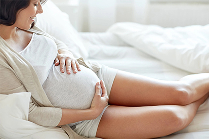
And the deciduous teeth contain stem cells
- Written by: Dr. Kouzi
Prospects for the use of stem cells from deciduous teeth in Regenerative Medicine of permanent teeth
Deciduous teeth and wisdom teeth are an important source of mesenchymal stem cells, similar to those from the umbilical cord. However, they have an important advantage: During human development,
part of the tooth is generated by the nervous system. This means that a percentage of the tooth stem cells retain neural stem cell characteristics, a property that stem cells from umbilical cord tissue do not possess. This specialized population constitutes approximately 5% of the stem cells of deciduous teeth and has already been used experimentally in the repair of peripheral nerves after injury. Although each deciduous tooth contains a smaller number of stem cells than umbilical cord tissue, their common origin with the nervous system makes them particularly valuable for applications in regenerative medicine of the nervous system.
The remaining cell populations of deciduous teeth are being tested in the creation of two individual tissues of permanent teeth, useful in their regeneration.
The tooth is a complex organ, with a particular shape and organization. Two separate lines of stem cells contribute to its creation, which play a crucial role in the formation of the dental roots and the surrounding alveolar bone. The replacement of lost teeth has been based for years on artificial solutions such as implants and dentures. Although effective, these options cannot fully reproducethe structure, function and physical feel of a real tooth. That is why scientists have been trying for decades to understand how teeth are formed with the goal of one day being able to regenerate them naturally.
The process of tooth formation is extremely complex and depends on the coordination of many different types of cells and tissues, such as the dental pulp, the enamel organ and the cells that form the jaw bone. The nervous system also participates in the formation and maintenance of teeth, and for this reason, in cases of aponeurosis,where the nerve of the tooth is destroyed, the tooth in the following years dies and falls out. Denervation relieved us of the pain, but led to the necrosis of the tooth.
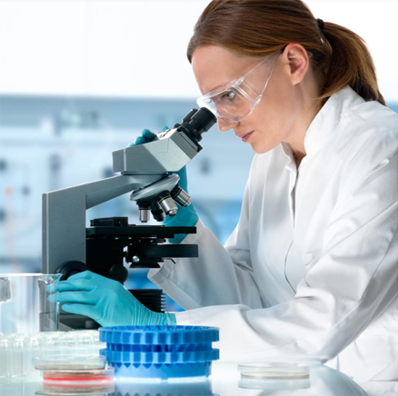
The research
To fill these knowledge gaps, a team led by Assistant Professor Mizuki Nagata from the Department of Periodontology, School of Medical and Dental Sciences, Tokyo Institute of Science, and Wanida Ono from the University of Texas Medical Science Center collaborated with researchers from the University of Michigan and other institutions. The team initially identified a population of mesenchymal stem cells in the pulp of deciduous teeth, which form the central substance of the tooth, and two distinct subpopulations within them, which follow different developmental pathways. The first population is closely associated with the formation of the tooth root, and the second contributes to the formation of the tissues that support it in the jaw. These discoveries provide a clearer picture of how teeth and the bone that supports them develop in the jaws. Mapping the role of these two lineages stem cells and the signals that guide them, researchers now have a stronger framework for understanding the formation of all the individual parts of the teeth.These findings pave the way for innovative regenerative therapies based on stem cells, which aim to regenerate the dental pulp and the survival of denervated teeth, periodontal tissues and jaw bones, replacing implants and artificial fillings. Since 2009, Biohellenika has offered the storage of all cell populations of deciduous teeth stem cells in their original form, and thisof neural origin. If the stem cells are placed in conditions of cell proliferation, the specialized population for neural tissue is lost and the baby tooth stem cells acquire the same uses as the umbilical cord tissue. This way of storage at Biohellenika ensures their maximum therapeutic potential for all future applications. Today, more than 950 clinical studies are underway in which mesenchymal stem cells are used, of which approximately 50 use baby tooth stem cells.
The American Academy of Pediatric Dentistry recommends the storage of baby tooth stem cells, due to their emerging uses in Regenerative Medicine. (Oral Health Policies, Dental Stem Cells, 2022).
Και τα νεογιλά δόντια περιέχουν βλαστοκύτταρα
- Written by: Dr. Kouzi
Προοπτικές της χρήσης των βλαστικών κυττάρων των νεογιλών δοντιών στην Αναγεννητική Ιατρική των μόνιμων δοντιών
Τα νεογιλά δόντια και οι σωφρονιστήρες αποτελούν μια σημαντική πηγή μεσεγχυματικών βλαστοκυττάρων, παρόμοιων με εκείνα του ομφάλιου λώρου. Ωστόσο διαθέτουν ένα σημαντικό πλεονέκτημα:
Κατά τη διάπλαση του ανθρώπου, μέρος του δοντιού δημιουργείται από το νευρικό σύστημα. Αυτό σημαίνει ότι ένα ποσοστό των βλαστοκυττάρων του δοντιού διατηρεί χαρακτηριστικά νευρικών βλαστοκυττάρων, ιδιότητα που δεν διαθέτουν τα βλαστοκύτταρα του ιστού του ομφαλίου λώρου.
Αυτός ο εξειδικευμένος πληθυσμός αποτελεί περίπου 5% των βλαστοκυττάρων των νεογιλών δοντιών και έχει ήδη χρησιμοποιηθεί πειραματικά στην αποκατάσταση περιφερικών νεύρων μετά από διατομή.
Παρότι κάθε νεογιλό δόντι περιέχει μικρότερο αριθμό βλαστοκυττάρων σε σχέση με τον ιστό του ομφαλίου λώρου, η κοινή τους προέλευση με το νευρικό σύστημα τα καθιστά ιδιαίτερα πολύτιμα για εφαρμογές στην αναγεννητική ιατρική του νευρικού συστήματος.
Οι υπόλοιποι κυτταρικοί πληθυσμοί των νεογιλών δοντιών δοκιμάζονται στη δημιουργία δύο επί μέρους ιστών των μόνιμων δοντιών, χρήσιμων στην αναγέννηση τους.
Το δόντι είναι ένα σύνθετο όργανο, με ιδιαίτερο σχήμα και οργάνωση. Στη δημιουργία του συμβάλλουν δύο χωριστές σειρές βλαστοκυττάρων που παίζουν καθοριστικό ρόλο στο σχηματισμό των οδοντικών καταβολών και του γύρω φατνιακού οστού.
Η αντικατάσταση των χαμένων δοντιών βασίζεται εδώ και χρόνια σε τεχνητές λύσεις όπως εμφυτεύματα και οδοντοστοιχίες. Αν και αποτελεσματικές, αυτές οι επιλογές δεν μπορούν να αναπαράγουν πλήρως τη δομή, τη λειτουργία και τη φυσική αίσθηση ενός πραγματικού δοντιού. Για αυτό το λόγο οι επιστήμονες προσπαθούν εδώ και δεκαετίες να κατανοήσουν πώς σχηματίζονται τα δόντια με στόχο να μπορέσουν κάποτε να τα αναγεννούν φυσικά.
Η διαδικασία διάπλασης των δοντιών είναι εξαιρετικά πολύπλοκη και εξαρτάται από τον συντονισμό πολλών διαφορετικών τύπων κυττάρων και ιστών, όπως είναι ο οδοντικός πολφός, το όργανο της αδαμαντίνης και τα κύτταρα που σχηματίζουν το οστό της γνάθου. Στη διάπλαση και διατήρηση των δοντιών συμμετέχει και το νευρικό σύστημα και για το λόγο αυτό στις περιπτώσεις της απονεύρωσης, όπου καταστρέφεται η νεύρωση του δοντιού, το δόντι στα επόμενα χρόνο νεκρώνεται και αποπίπτει. Η απονεύρωση μας απάλλαξε από τον πόνο, αλλά οδήγησε στη νέκρωση του δοντιού.
Οι έρευνες
Για να καλύψει αυτά τα κενά γνώσης, μια ομάδα με επικεφαλής την επίκουρη καθηγήτρια Μιζούκι Ναγκάτα από το Τμήμα Περιοδοντολογίας της Σχολής Ιατρικών και Οδοντιατρικών Επιστημών του Ινστιτούτου Επιστήμης του Τόκιο και τη Γουανίντα Όνο από το Κέντρο Ιατρικής Επιστήμης του Πανεπιστημίου του Τέξας συνεργάστηκαν με ερευνητές από το Πανεπιστήμιο του Μίσιγκαν και άλλα ιδρύματα.
Η ομάδα αρχικά εντόπισε μέσα στον πολφό των νεογιλών δοντιών έναν πληθυσμό μεσεγχυματικών βλαστοκυττάρων, τα οποία δημιουργούν την κεντρική ουσία του δοντιού και μεταξύ αυτών δύο διαφορετικούς υποπληθυσμούς, οι οποίοι ακολουθούν διαφορετική αναπτυξιακή πορεία. Ο πρώτος πληθυσμός συνδέεται στενά με τον σχηματισμό της ρίζας του δοντιού και ο δεύτερος συμβάλλει στο σχηματισμό των ιστών που το στηρίζουν στη γνάθο.
Οι ανακαλύψεις αυτές προσφέρουν μια πιο καθαρή εικόνα για το πώς αναπτύσσονται τα δόντια και το οστό που τα στηρίζει μέσα στις γνάθους. Χαρτογραφώντας το ρόλο αυτών των δύο σειρών βλαστοκυττάρων και τα σήματα που τις καθοδηγούν, οι ερευνητές διαθέτουν πλέον ένα ισχυρότερο πλαίσιο κατανόησης του σχηματισμού όλων των επί μέρους τμημάτων των δοντιών.
Τα ευρήματα αυτά ανοίγουν το δρόμο για καινοτόμες αναγεννητικές θεραπείες βασισμένες σε βλαστοκύτταρα, οι οποίες αποσκοπούν στην αναγέννηση του οδοντικού πολφού και την επιβίωση των απονευρωμένων δοντιών, των περιοδοντικών ιστών και των οστών των γνάθων, αντικαθιστώντας τα εμφυτεύματα και τα τεχνητά σφραγίσματα. Η Biohellenika από το 2009 προσφέρει τη φύλαξη όλων των κυτταρικών πληθυσμών των βλαστοκυττάρων των νεογιλών δοντιών στην αρχική τους μορφή, και αυτόν της νευρικής προέλευσης. Εάν τα βλαστοκύτταρα τεθούν σε συνθήκες κυτταρικού πολλαπλασιασμού χάνεται ο εξειδικευμένος για τον νευρικό ιστό πληθυσμός και τα βλαστοκύτταρα των νεογιλών δοντιών αποκτούν τις ίδιες χρήσεις με τον ιστό του ομφαλίου λώρου. Με τον τρόπο αυτό της φύλαξης στην Biohellenika εξασφαλίζεται το μέγιστο θεραπευτικό δυναμικό τους για όλες τις μελλοντικές εφαρμογές.
Σήμερα βρίσκονται σε εξέλιξη περισσότερες από 950 κλινικές μελέτες στις οποίες χρησιμοποιούνται μεσεγχυματικά βλαστοκύτταρα, εκ των οποίων περίπου 50 χρησιμοποιούν βλαστοκύτταρα νεογιλών δοντιών.
Η Αμερικανική Ακαδημία Παιδιατρικής Οδοντιατρικής συνιστά την αποθήκευση βλαστοκυττάρων νεογιλών δοντιών, λόγω διαφαινόμενων χρήσεων τους στην Αναγεννητική ιατρική. (Oral Health Policies, Dental Stem Cells, 2022).
Αποτελεσματικές κυτταρικές θεραπείες για το AIDS
- Written by: Dr. Kouzi
Νέα προοπτική για τη θεραπεία της μόλυνσης από τον ιό HIV
Στο πρόσφατο συνέδριο της Αμερικανικής εταιρείας Κυτταρικής και Γονιδιακής θεραπείας που έγινε στην Βοστώνη των ΗΠΑ 11-15 Μαΐου 2026 ανακοινώθηκαν τα αποτελέσματα μικρής κλινικής μελέτης που αφορούσε τη θεραπεία του συνδρόμου της επίκτητης ανοσοανεπάρκειας (AIDS) που προκαλείται από την μόλυνση από τον ιό HIV με τη χρήση των CAR -T κυττάρων. Τα κύτταρα αυτά στοχεύουν και καταργούν τις θέσεις σύνδεσης του ιού HIV επάνω στα Τ λεμφοκύτταρα, η κατάληψη των οποίων από τον ιό οδηγεί στην εξασθένηση του ανοσοποιητικού συστήματος του ασθενή. Σήμερα 41 εκατ. άνθρωποι σε όλο τον κόσμο πάσχουν από AIDS, οι οποίοι λαμβάνουν εφ’ όρου ζωής θεραπεία και διατηρούν μία φυσιολογική ζωή.
Σε λίγους ασθενείς έχει γίνει μεταμόσχευση μυελού των οστών από δότες οι οποίοι εκτός από την ιστοσυμβατότητα διαθέτουν και το γονίδιο το οποίο προστατεύει τα λεμφοκύτταρα από την προσκόλληση του ιού HIV σε αυτά. Η χρήση των CAR T θα χορηγηθεί στη συνέχεια σε μία δόση και σε μεγαλύτερη ομάδα ασθενών.
Στην κλινική μελέτη μετέχουν τα Πανεπιστήμια San Francisco της California, Davis και Case Western Reserve University Hospital. Από τον έλεγχο των ασθενών μετά τη χορήγηση των CAR T βρέθηκε ότι μερικοί διατήρησαν μη ανιχνεύσιμα επίπεδα του ιού, ενώ άλλοι πολύ χαμηλά επίπεδα μέχρι δύο χρόνια από τη χορήγηση, όσο διήρκεσε η παρακολούθηση των ασθενών. Στους ασθενείς που παρατηρήθηκε η πλέον μακρόχρονη ύφεση η θεραπεία είχε εφαρμοστεί σε πολύ σύντομο χρόνο μετά τη διάγνωση. Οι επιπλοκές είναι ελάχιστες σε σχέση με άλλες χρήσεις των CAR-T για τη θεραπεία κακοήθων ασθενειών.
Τα CAR-T κύτταρα δημιουργούνται από τα κύτταρα του ανοσοποιητικού και μία πηγή λήψης αυτών είναι το ομφαλοπλακουντιακό αίμα, το οποίο διαθέτει τα πλέον υγιή και νεαρά κύτταρα. Το ομφαλοπλακουντιακό αίμα περιέχει και άλλα είδη κυττάρων του ανοσοποιητικού, τα οποία δοκιμάζονται κλινικά και προσφέρουν ακόμα πιο αποτελεσματικές θεραπείες για κακοήθεις αυτοάνοσες παθήσεις.


![]()


Biohellenika Stem Cells Bank
Welcome to the official website of the most prominent, accredited, licensed and well-equipped stem cell bank. Visit our offices and accredited laboratories


Families
trusted us
The first
therapeutic application of stem cells in Greece was made by us
Years
we are the first in quality and stem cells applications
Publications
in scientific community
Patents
Worldwide